

Golden 10 years thorax sagittal

The ribs of this patient are parallel. Does the cat have the costal alignment of the dog? How do they differ? Subscribe to pro for the answer!
Golden 10 years thorax sx
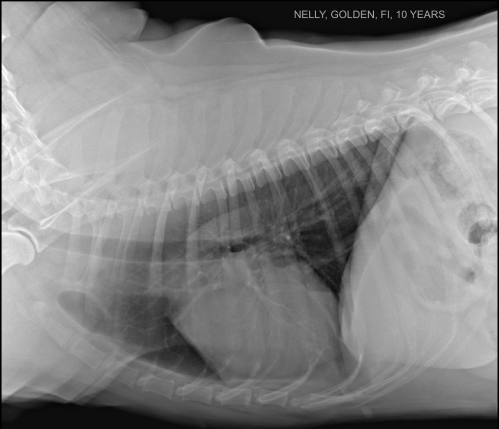
What is the thin, soft tissue opaque line superimposed on the caudal aspect of the cardiac silhouette? Has this patient a pleural effusion?
Golden 10 years thorax dx
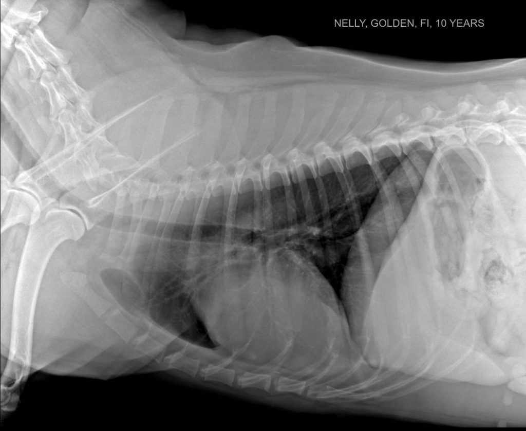
It is common in Golden Retrievers to have an unfolding dorsal tracheal membrane visible in the cervical trachea. This finding has not been confused with a collapsing trachea syndrome.
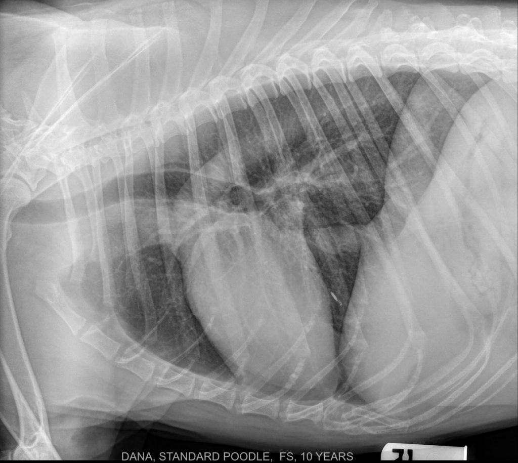
Standard Poodle 10 Years Thorax Sagittal
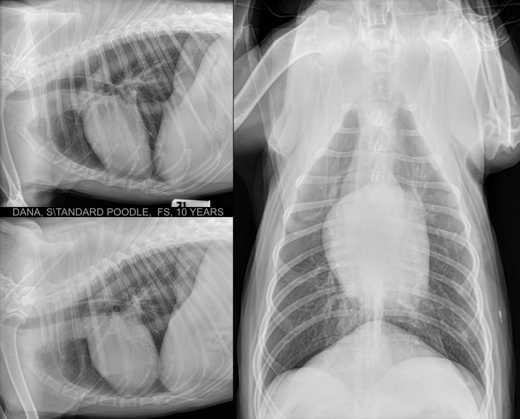
A drop of gas is in the patient’s esophagus on both views. This finding could be because the patient was panting during the procedure or could be a sign of mild esophagitis. Which is the other common cause for detecting a gas-dilated esophagus on a thoracic radiograph? Please, correlate the radiographic findings with the clinical […]
Standard Poodle 10 Years Thorax Sx
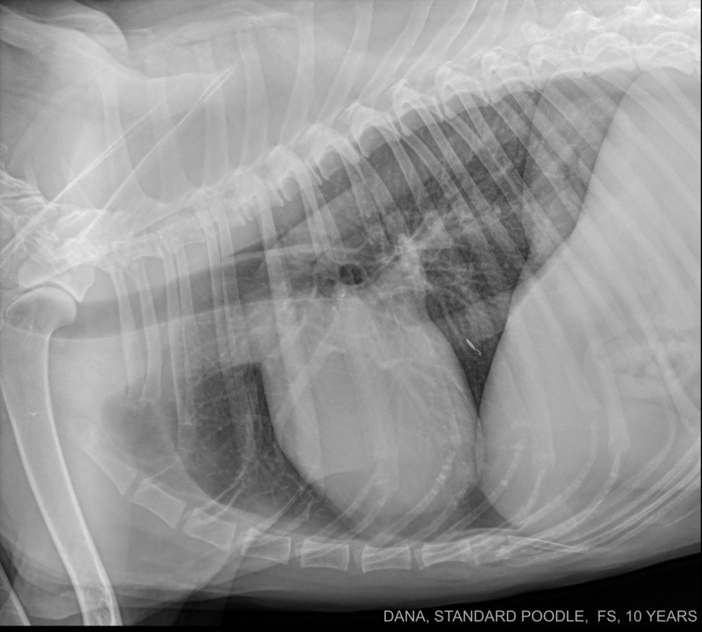
What structure is the faintly visible, tubular-shaped, soft tissue opacity visible in the caudal portion of the thorax, ventral to the aorta, and dorsal to the caudal vena cava?
Standard Poodle 10 Years Thorax Dx
Funny where a microchip can migrate!
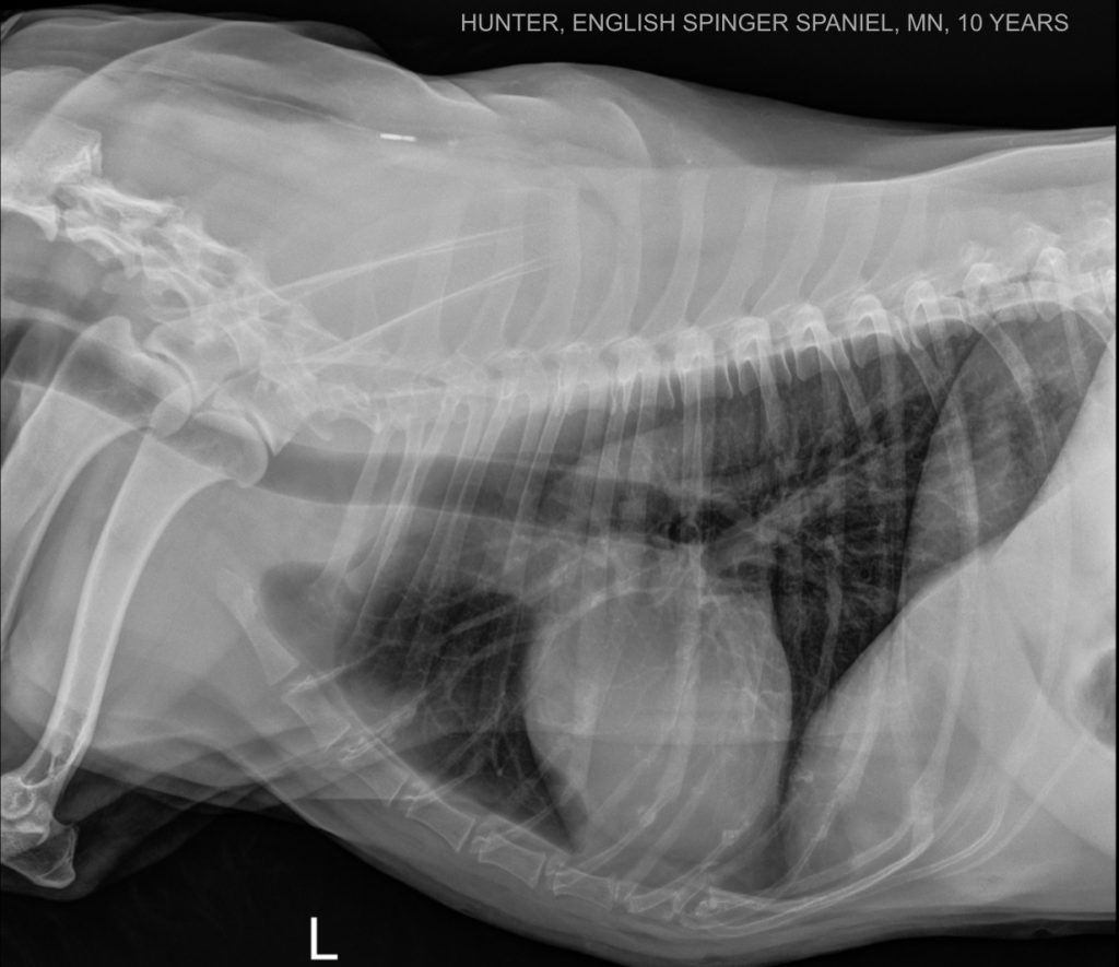
English Springer Spaniel 10 Years Thorax Sagittal
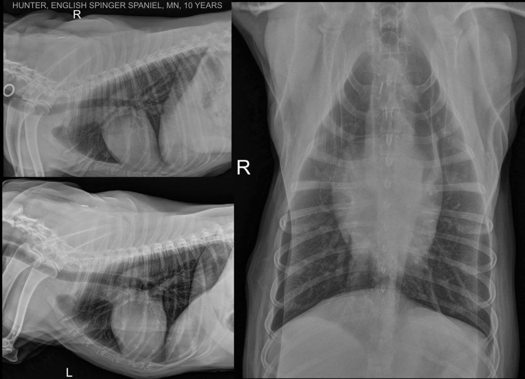
What are the two radiopaque lines that run nearly parallel to the thoracic walls in the VD views, mimicking a mild pneumothorax? Subscribe to pro for the answer!
English Springer Spaniel 10 Years Thorax Sx
The front limbs should be perfectly aligned, and minimal rotation should be present. It is impressive how much the shape of the cardiac silhouette changes in the different projections! Do you want to know why? Subscribe to pro!
English Springer Spaniel 10 Years Thorax Dx
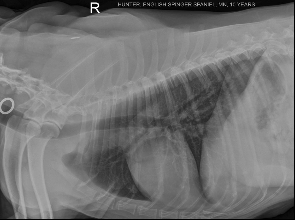
A metallic ring is seen superimposed on the cervical trachea. Remember to remove every radiopaque object from the primary beam!