

Pinscher 7 Years Thorax Sx
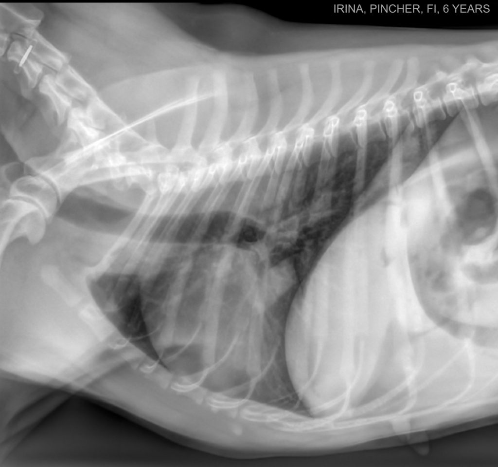
What is not within normal limits in this image? You can also check the other views.
Pinscher 7 Years Thorax Dx
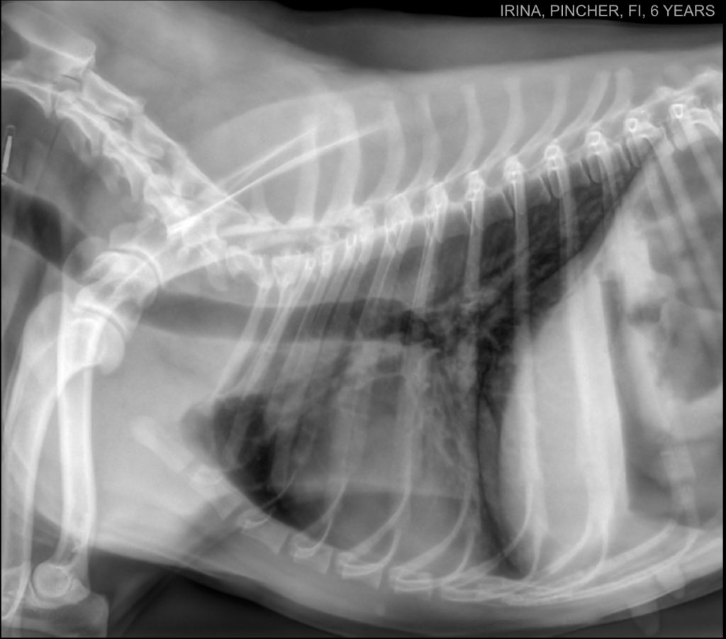
On lateral views, the cardiac silhouette is nearly occupying all the thorax. If you have to stage the patient for lung metastasis, consider obtaining one lateral view, one DV, and one VD.
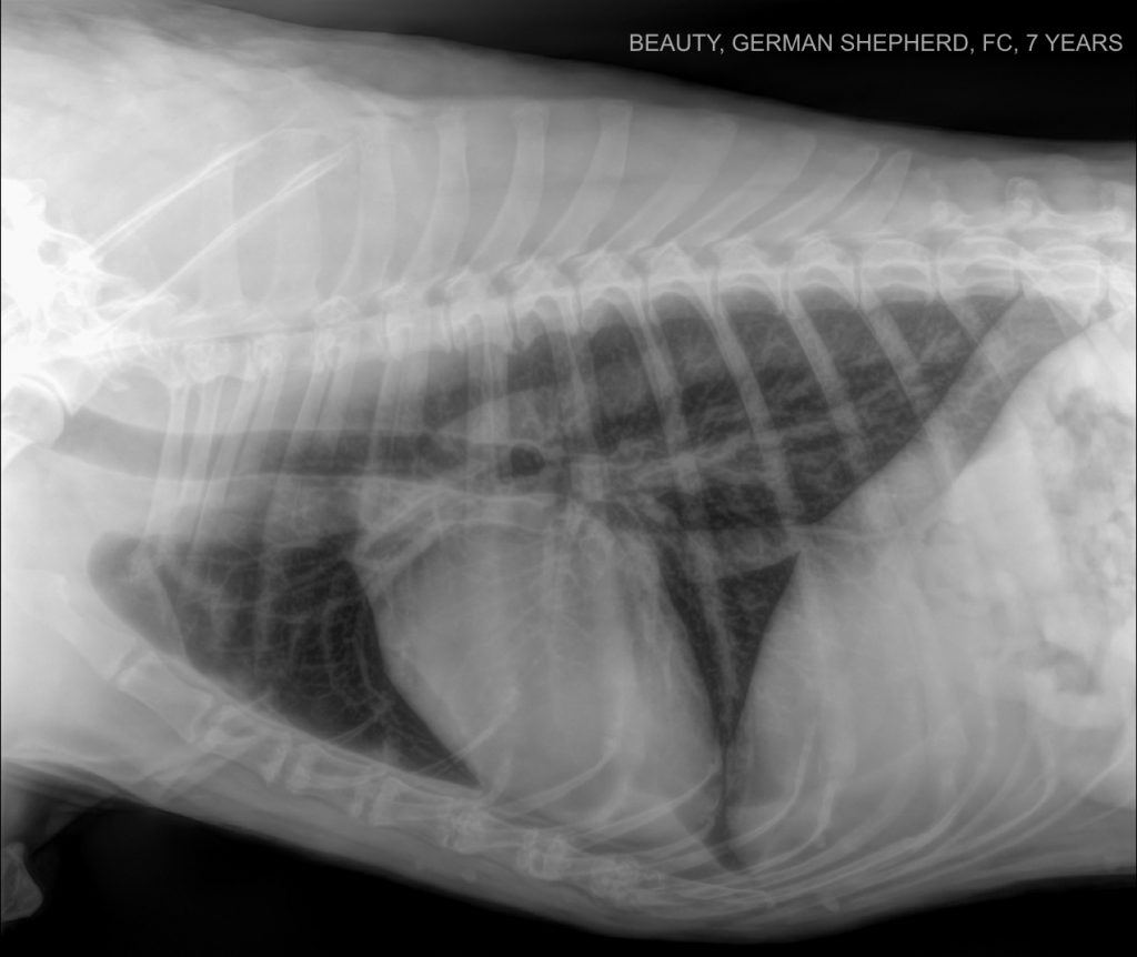
German Shepherd 7 Years Thorax Sagittal
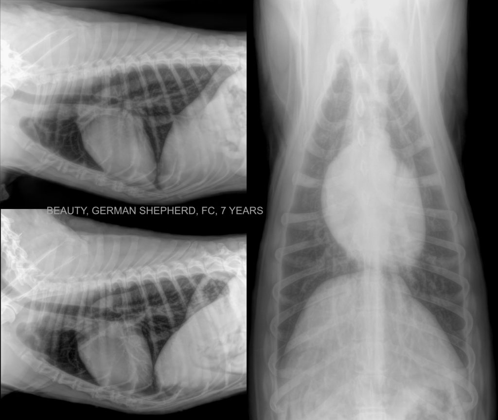
What is the oblique line visible in the DV view, starting cranial to the first rib to the right and leading toward the left in the areas of the cranial lung lobes? Subscribe to pro for the answer!
German Shepherd 7 Years Thorax Sx
There is a mild rotation in this projection, sorry!
German Shepherd 7 Years Thorax Dx
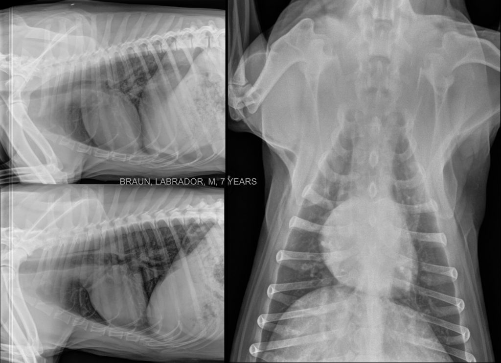
Look at the degenerative changes in the sternum of this patient. These degenerative changes are common, especially in large-breed dogs, but the clinical significance is not clear.
Labrador 7 Years Thorax Sagittal
The caudal tips of the lungs are missing in the sagittal image. In this case, the image should have been centered more caudally.
Labrador 7 Years Thorax Sx
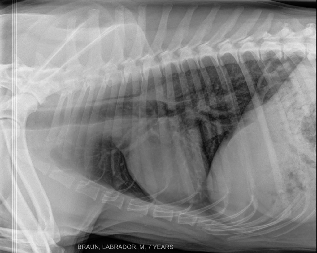
There are digital artefacts in the cranial aspect of the image (three, metallic radiopaque, parallel lines) due to the cassette developer in a computed indirect digital system.
Labrador 7 Years Thorax Dx
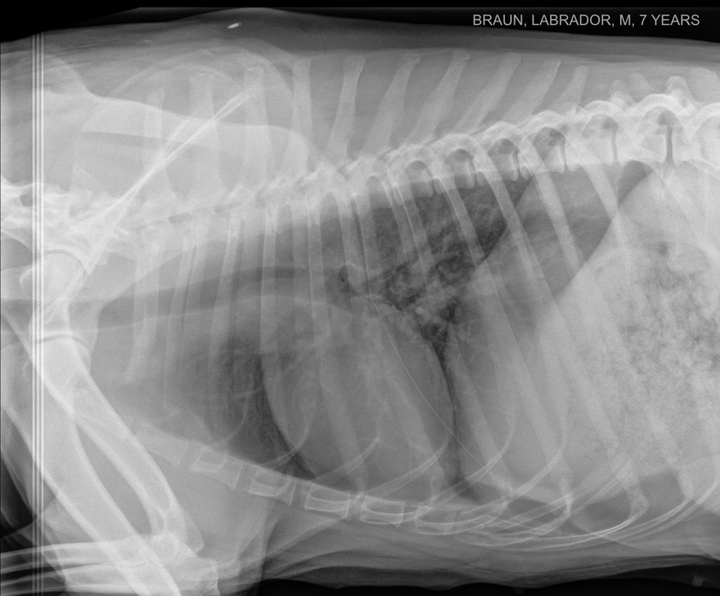
This projection has a mild rotation, more severe at the caudal aspect of the thorax, sorry!